As global food requirements intensify and farming moves decisively toward sustainable and efficient models, establishing precise, forward-looking strategies to protect livestock health is increasingly critical. Within the egg and hatchery domain, hyperspectral imaging (HSI) has established itself as a sophisticated, non-invasive modality for swift, reliable egg quality evaluation, including the detection of embryonic death in chicks. Yet, steep costs and operational barriers relative to conventional RGB approaches present considerable barriers to HSI’s wide-scale uptake. Circumventing these constraints and enabling the broader promise of HSI, the reconstruction of hyperspectral data from ordinary RGB captures is a compelling answer. The current work focuses on producing hyperspectral imagery from RGB inputs to achieve non-destructive, early-stage identification of chick embryo mortality. An initial comparison was made among several image reconstruction frameworks—HRNET, MST++, Restormer, and EDSR—for generating hyperspectral representations of eggs at an early incubation phase. Following this, the reconstructed spectral information was used as input for XGBoost and Random Forest models to separate eggs with living embryos from those with deceased embryos. HRNET outperformed the alternatives in reconstruction quality, recording an MRAE of 0.0955, an RMSE of 0.0159, and a PSNR of 36.79 dB. This work promotes the concept that integrating imaging platforms with intelligent sensing and analytical processing can raise automation levels, strengthen biosecurity measures, and streamline resource use in pursuit of sustainable agriculture 4.0.
Rising worldwide per capita egg intake is creating a clear imperative for the rollout of modern automated sorting and grading infrastructure capable of meeting expanding market requirements. A combination of Industry 4.0 thinking and mounting international concern for animal welfare and ecological footprints has brought non-destructive, non-invasive methodologies to the forefront in egg and hatchery operations [1, 2]. HSI functions as a swift, precise, non-destructive optical sensor technology, sampling the electromagnetic spectrum across a dense set of narrow, contiguous wavebands and providing rich insight into the multifaceted character of egg traits [3]. By enabling the observation of the spatial patterns of quality characteristics, HSI provides a versatile means for real-time oversight and regulation across numerous settings [4-6]. A body of literature suggests that HSI might radically alter egg and hatchery practices given its ability to pick up vital indicators such as sex, fertility status, and embryo death [7, 8]. When smart sensing is coupled with HSI, hatchery operators are empowered to keep an integrated watch over environmental parameters and execute knowledge-driven interventions—varying incubation settings, guiding selective breeding choices, or spotting early fatalities—strengthening overall biosecurity [3, 9]. The phenomenon of embryos perishing within their shells, known as embryonic mortality, has significant implications for economic returns, output capacity, genetic health, animal care standards, and environmental accountability [10]. Causes range from hereditary errors and external stress agents to infectious diseases [11]. Although embryonic growth commences within a 24–36-hour (hr) window following incubation onset, chicken eggs need roughly 21 days to reach hatching readiness [12]. Time-honored methods for embryo assessment rely heavily on manual effort, are subjective, and lack throughput. Alternatives such as HSI, along with other non-destructive tools such as photoplethysmography (PPG) and thermal imaging, have emerged as efficient solutions [13-15]. These state-of-the-art techniques provide early, reliable detection of chick embryo deaths, facilitating swift removal of unviable eggs, thereby elevating hatch success and overall production efficiency in poultry systems.
Regardless of the merits of HSI, implementing it in practice is impeded by financial outlay, the enormous data footprint, and lagging data capture rates [16]. The high price of hyperspectral cameras and supporting components acts as a brake on adoption, particularly for institutions managing tight budgets. Moreover, the sheer volume and complexity of the data necessitate substantial computational resources [17]. A further key weakness of HSI in egg-based studies is its reliance on specific lighting geometries to expose internal characteristics, which could interfere with the egg’s biology and threaten continued embryo growth [18]. As such, to transcend present-day boundaries surrounding HSI, computational approaches such as deriving hyperspectral information from standard RGB photos could open new avenues for scientific inquiry, industrial use, and policy formulation.
Hyperspectral image reconstruction centres on extracting details across a succession of contiguous wavebands from standard tri-band RGB captures. A central merit of such reconstruction is its reach, given that it makes expensive, dedicated HSI hardware unnecessary. The accelerating interest in this area is spurred by the straightforward, near-universal availability of RGB imaging devices, characterized by low sensor costs and light data storage demands [16, 19]. All the same, the reconstruction process introduces considerable hurdles, including mapping ambiguities, scene-dependent spectral variation, trade-offs between spatial and spectral accuracy, noise, and visual imperfections in source RGB imagery [20]. An array of tactics leaning on advanced network architectures, synthetic data expansion, transfer learning, and denoising methods has been documented as a means of tackling these obstacles [21]. A well-chosen reconstruction model could substantially benefit egg and hatchery enterprises by enabling tasks spanning grading and sorting, quality surveillance, and anticipatory identification of fertility and sex, while leaving egg biology undisturbed compared to traditional HSI workflows.
Despite strong upside potential, only a limited number of studies have examined hyperspectral reconstruction of biological and agricultural specimens [22, 23]. Pioneering work involved reconstructing hyperspectral data from learned manifolds rather than directly from RGB, under the hypothesis that hyperspectral pixels lie on low-dimensional manifold spaces [21]. Beginning with the New Trends in Image Restoration and Enhancement (NTIRE) challenge on spectral recovery from RGB, numerous convolutional neural network (CNN), attention-oriented, and transformer-powered architectures have been proposed [24]. These designs, though still requiring comprehensive validation, hold promise for accurately rebuilding representations of elaborate biological and agricultural items. Recently, Yang et al. [22] applied a pretrained MST++ pipeline to construct inversion frameworks for physiological rice indicators, achieving an R² of 0.40 in forecasting SPAD values. Zhao et al. [23] developed a robust HSCNN-R model for hyperspectral recovery from RGB inputs to estimate the soluble solid content (SSC) of tomatoes, achieving an R² of 0.51. Somewhat later, Ahmed et al. [25] examined the HSCNN-D architecture, returning an R²p of 0.69 for predicting sweet potato SSC. Elsewhere, researchers [26] weighed three hyperspectral recovery algorithms—HSCNN-D, MST++, and HRNET—for predicting sweet potato dry matter content (DMC). The HRNET technique yielded spectra that closely matched reference measurements, yielding an R²p of 0.88. These inquiries highlighted the feasibility of image recovery for evaluating the quality of agricultural and biological samples. Extending that foundational work, this paper constitutes the earliest attempt at egg hyperspectral image reconstruction aimed at advancing egg and hatchery science. Specifically, the objective of this study is to deploy leading-edge reconstruction models, namely HRNET, MST++, Restormer, and EDSR, to regenerate hyperspectral information from RGB captures for the early forecasting of chick embryo mortality.
The research adhered to the animal experimentation procedures (protocol #: 22,224) sanctioned by the Office of the Vice-Chancellor for Research IACUC and the IACUC online protocols at the University of Illinois Urbana-Champaign (UIUC). From the UIUC poultry facility, 102 white-shell chicken eggs were gathered. Every mother hen was of the White Leghorn variety, aged around 29 weeks. It bears mentioning that only 9 eggs from the total of 102 displayed embryonic death, with causes linked to uncontrollable elements like inherited abnormalities and external stress factors. The eggs were incubated in a single-stage commercial unit (Natureform Inc., Jacksonville, FL, USA) set to 99.5° F, with 85% relative humidity and an automatic hourly turn of 45°. At the end of the full incubation cycle, the egg contents were exposed and examined to determine whether the embryo was alive or dead.
A benchtop line-scan HSI apparatus (Resonon Inc., Bozeman, MT, USA) served as the platform for collecting hyperspectral imagery (Figure 1). Core components comprised a 900-pixel line-scanner detector (Pika L, Resonon, Inc.) operating across the 400–1000 nm window paired with a 23 mm focal length optic, a tungsten-halogen broad-spectrum illuminator, a linear movable translation platform, and a connected computing unit. With ambient lighting extinguished, all captures were executed through Spectronon Pro software (Resonon Inc., Bozeman, MT, USA) running on the linked computer. Every individual egg was set upright in a dark-colored cradle, air cell facing upward, while the illumination source was stationed immediately below it. The light source warm-up lasted 30 minutes before any imaging commenced, ensuring consistent radiant output throughout the experimental timeline. To guarantee reproducibility in image quality, both the lens-to-specimen distance (20 cm) and all capture parameters configured in Spectronon Pro—specifically “stage” (scan rate: 0.07 cm/s, homing rate: 1.5625 cm/s, jog rate: 0.07 cm/s) together with “imager” (frame frequency: 9.9 fps, integration window: 100 ms, gain: 0)—were kept unchanged throughout. Before scanning, each tray holding 30 eggs was removed from the incubator and kept warm on an electric blanket (90–95° F) while the entire grouping was imaged. The same procedure was followed to acquire hyperspectral images on incubation days 2, 3, and 4. This yielded 306 images (102 × 3 days) ready for downstream processing. One key observation is that light incident on an egg does not follow a straight path; rather, it disperses into multiple trajectories, altering both direction and amplitude. Such scattering effects cause portions of the light to be absorbed or reflected by the egg structure, making it difficult to isolate purely transmitted light. For this reason, digital number values [27] (alternatively called pixel intensity) were adopted for subsequent analysis rather than derived quantities such as transmittance or absorbance.
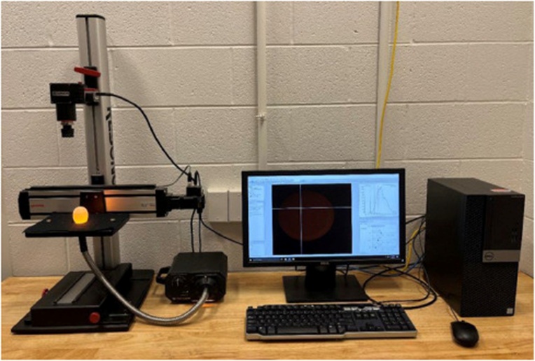
Figure 1. The line-scan hyperspectral imaging system for non-destructive egg analysis.
Extracting spectral content from hyperspectral images hinges critically on an initial segmentation operation [28]. The task involves demarcating regions of interest (ROIs) within the examined specimen by constructing a binary mask that isolates them from the rest of the scene. Here, that mask was derived at the 700 nm band—selected for exhibiting the strongest contrast differential between egg and backdrop—with a cutoff set at 0.02. The resulting mask then defined the ROI from which spectral data were subsequently retrieved. One averaged spectral profile was produced per hyperspectral image by averaging the spectra of all pixels within each ROI.
The reconstruction frameworks expect RGB images at their input and rely on hypercubes as supervisory labels. A complication arises because the Resonon HSI setup does not simultaneously deliver RGB renditions alongside the hyperspectral captures. To resolve this, synthetic pseudo-RGB images were assembled by assigning the 600 nm band to the red channel, the 549 nm band to the green channel, and the 450 nm band to the blue channel. These assembled images were stored as JPEG (Joint Photographic Experts Group) files (Figure 2). Subsequently, the pseudo-RGB images served as the source material for the reconstruction algorithms.
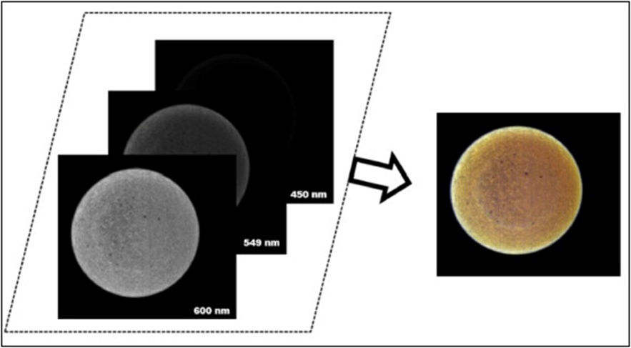
Figure 2. Generation of pseudo-RGB images combining 600, 549, and 450 nm bands.
Forming the reference ground-truth hypercubes involved selecting, at random, a set of non-collinear wavelengths spanning the visible to near-infrared (VIS-NIR) region from 400 to 1000 nm. This random selection yielded hypercubes with dimensions 800 × 900 × λ, where λ denotes the number of wavelengths selected. These hypercubes were subsequently employed as the labeled training targets during the learning phase of the reconstruction algorithms.
Four distinct architectural paradigms were selected to deliver a balanced assessment of reconstruction capability (Figures 3a-3d). EDSR is rooted in a CNN framework; Restormer and MST++ lean heavily on attention mechanisms within their designs. HRNet primarily relies on CNN principles while integrating selective self-attention components. Convolutional and attention-based networks emphasize the contrasting characteristics of visual inputs. Attention-oriented models behave like low-pass filters, whereas convolution-centric models resemble high-pass filters [29]. By systematically comparing these architecturally divergent approaches, this investigation aims to shed light on viable pathways forward for spectral reconstruction specific to egg analysis.
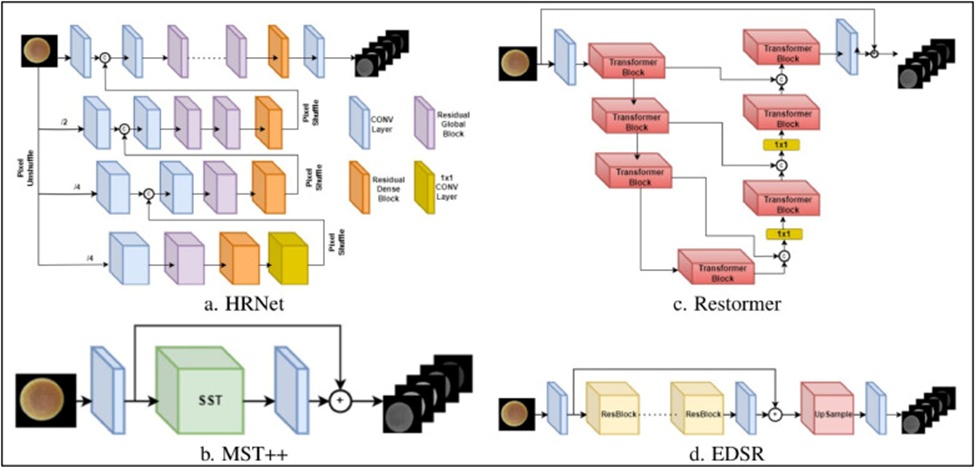
Figure 3. Architectures of HSI reconstruction models. All the reconstruction models take RGB as input and provide HSI (λ bands) as output.
HRNet arranges input imagery into a series of hierarchical levels through successive twofold reductions in resolution [23]. HRNet integrates PixelUnshuffle as its downscaling mechanism, achieving reduced resolution without introducing new learnable parameters. Each hierarchical tier is processed jointly with the upsampled representations flowing upward from the tier directly beneath it. PixelShuffle handles the upscaling operation. This sequential refinement climbs until reaching the uppermost level, which delivers the reconstructed spectral output. Rather than employing fixed interpolation schemes, the upsampling transforms are learned during training—the overarching design philosophy of HRNet is to preserve information fidelity throughout successive resolution reductions.
MST++ is built on a Single-Stage spectral-wise Transformer (SST) architecture, composed of multiple mask-steered spectral-wise multi-head self-attention blocks [30]. The incorporated masks direct computational focus to the most salient aspects of the hyperspectral representation, enabling MST++ to capture extended-range intra-spectral relationships in hyperspectral datasets faithfully.
Restormer, first devised as a transformer-driven general image restoration engine, has been successfully extended to the problem of hyperspectral reconstruction [31]. Its distinctive design applies self-attention computations across the channel axis rather than over spatial coordinates, making it particularly adept at uncovering interdependencies spanning different spectral bands. Each transformer block in Restormer contains a multi-Dconv head transposed attention module that orchestrates query-key feature interactions in the channel domain. Complementing this, the architecture incorporates a gated feed-forward structure that selectively permits only the most information-bearing representations to advance deeper into the network.
Rooted in the SRResNet lineage [32], EDSR [33] chains together a succession of residual blocks for hyperspectral recovery. Its designers recognized that removing the batch normalization layers from these residual blocks yields negligible reductions in reconstruction fidelity while substantially reducing memory requirements. Exploiting this insight, EDSR packs more residual blocks into an equivalent memory budget, thereby boosting representational depth without sacrificing computational feasibility.
The MRAE metric computes the relative magnitude of absolute discrepancies between model predictions and the ground truth (GT) targets. Because it captures the mean relative deviation rather than raw differences, MRAE offers the advantage of scale invariance, unlike metrics that factor in only absolute departures.
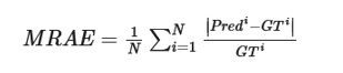 (1)
(1)
For RMSE, each prediction’s deviation from its corresponding GT value gets squared, summed, and then square-rooted. Squaring the individual error terms amplifies the influence of large deviations, making RMSE particularly responsive to outliers and well-suited to applications where such extreme departures should be given greater weight.
 (2)
(2)
PSNR serves as an indicator of how faithfully a reconstructed image retains its original signal content, defined as the ratio of the maximum signal strength achievable to the contaminating noise level.
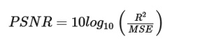 (3)
(3)
Given the limited size of the spectral dataset, classification was performed using XGBoost [34] and Random Forest [35], with model performance evaluated via stratified 5-fold cross-validation. XGBoost belongs to the class of boosting algorithms: it builds an ensemble in a stepwise fashion, where each successive weak learner is trained to correct the errors left by earlier learners. This sequential bias-toward-error mechanism renders XGBoost particularly valuable when overfitting poses a threat. By contrast, a Random Forest constructs an ensemble comprising many individual decision trees, each grown from a randomly drawn feature subset and trained on a bootstrapped sample of the data. Classification decisions are determined by aggregating votes across all trees, with the most-voted class selected as the output. Accuracy, precision, recall, and the F1 score served as the assessment criteria for the classifiers deployed herein (4), (5), (6), (7).
 (4)
(4)
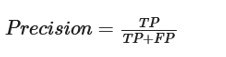 (5)
(5)
 (6)
(6)
 (7)
(7)
Where TP indicates true positive, FP false positive, TN true negative, and FN false negative.
All numerical experiments were executed on Google Colab Pro, a cloud-hosted environment. The allocated virtual machine contained a dual-core Intel(R) Xeon(R) CPU operating at 2.30 GHz, 25 GB of RAM with extended runtime, plus an NVIDIA Tesla P100 GPU with 16 GB of VRAM. Programming was performed in Python 3.9. Open-source libraries—namely, Scikit-learn, PyTorch, and OpenCV—were leveraged to reconstruct and evaluate the models.
Preparing the input RGB stream and the label hyperspectral cubes constituted the first step of the training pipeline. For the label side, 10 discrete spectral channels were retained from the VIS-NIR domain (400–1000 nm): 520, 583, 619, 655, 700, 739, 780, 837, 870, and 903 nm. Spectral inspection guided this choice, as these wavelengths showed clear differentiation between viable and non-viable embryos. The selected bands track physiological phenomena such as hematogenesis, yolk resorption, and fluctuations in water, protein, and lipid reservoirs during embryogenesis [36-38]. Shifts in these biochemical markers reflect the evolving metabolic exchange between the embryo and its egg environment [39, 40]. A feature-reduction procedure was applied to remove collinear redundancy from the original hyperspectral cubes [41]. Once pruned to the 10 selected wavelengths, the hyperspectral data served as ground-truth supervision, while the matching RGB renderings were fed as model inputs. From the overall pool of 306 images, a random split allocated 80% to training, 10% to validation, and the remaining 10% to testing. The Adam optimizer drove the parameter updates, with decay rates β1 = 0.9 (adjusted to 0.5 for HRNet) and β2 = 0.99. Training proceeded with a batch size of 1, a spatial patch size of 128, and a stride value of 8. The learning rate was initialized at 1e-4 and reduced exponentially after each epoch to improve convergence.
Having fixed these hyperparameters, each candidate architecture was trained for 200 epochs, with 1000 iterations per epoch. The MRAE loss function was adopted for its ability to uniformly assess spectral bands across illumination conditions and its documented resistance to the pull of noisy data, a point noted by Shi et al. [42]. Moreover, earlier work by Zhao et al. [23] demonstrated that reliance on the MRAE loss facilitates faster convergence, often within fewer epochs. Over the course of training, the loss trace flattened after a certain number of epochs, signaling that further improvement had essentially halted. Crucially, model snapshots were retrieved for downstream testing from the point just before this plateau materialized. This optimal moment occurred at epochs 189 for HRNET, 175 for MST++, 180 for Restormer, and 195 for EDSR. These saved checkpoints were then evaluated against the held-out validation and test partitions. A summary of the resulting metrics appears in Table 1.
Table 1. Evaluation metrics of the hyperspectral image reconstruction models on the validation and test sets. Bold indicates the best performance in each metric.
Methods | PSNR (dB) | RMSE (test) | MRAE (test) | RMSE (val) | MRAE (val) |
HRNet | 36.79 | 0.0159 | 0.0955 | 0.0151 | 0.0936 |
MST++ | 13.51 | 0.0377 | 0.2615 | 0.0378 | 0.2615 |
Restormer | 35.32 | 0.0188 | 0.1111 | 0.0188 | 0.1111 |
EDSR | 35.70 | 0.0178 | 0.1136 | 0.0178 | 0.1136 |
Among the reconstruction pipelines benchmarked, HRNet achieved the best overall metrics, with the lowest recorded MRAE of 0.0936, minimal RMSE of 0.0151, and peak PSNR of 36.79 dB. This fidelity came at a computational price, however: HRNet was the most resource-intensive configuration, requiring 40.5 hours and 9.49 gigaflops (G) of computation. At the opposite end of the efficiency spectrum, MST++ posted the smallest resource footprint (0.15 G) and ran for only 12.5 hours, yet its PSNR performance suffered substantially, attaining merely 13.51 dB. Restormer occupied an intermediate position, consuming 5.39 G and finishing in 37 hours while delivering a competitive PSNR of 35.32 dB. EDSR, meanwhile, surpassed both MST++ and Restormer on PSNR, achieving 35.70 dB, despite a heavy computational demand of 9.17 G.
After successful model execution, a visual comparison was performed to assess how closely the reconstructed hyperspectral outputs matched the reference ground-truth images. Both sets, across several representative spectral bands, are laid out in Figure 4 for inspection. A visual inspection of Figure 4 confirms that the reconstructed results closely resemble their ground-truth counterparts. The images produced by HRNet and Restormer, in particular, displayed striking visual fidelity. Yet, upon closer examination, Restormer outputs revealed subtle intensity irregularities, most evident at the 739 and 780 nm bands. EDSR-generated images exhibited a noticeable contrast offset relative to the ground truth in the 700 nm channel, as shown in Figure 4, while maintaining good visual agreement elsewhere. On the other hand, the MST++ reconstructions stood out for clearly visible pixel-intensity discrepancies relative to the ground truth; this method struggled markedly to faithfully reproduce intensities, especially in the spectral interval spanning 583-700 nm, as highlighted in Figure 4.
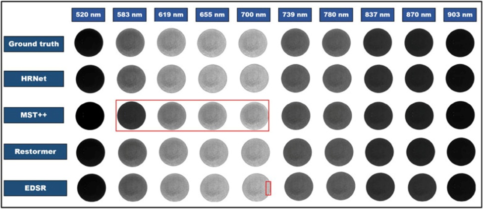
Figure 4. Ground-truth and reconstructed images. The red area represents the inconsistencies in the reconstructed pixels. MST++ showed the greatest inconsistencies in reconstructing the 583, 619, 655, and 700 nm bands.
Once the image reconstruction stage was completed, spectra were drawn from the reconstructed outputs, and their fidelity was systematically evaluated. Figures 5a-5d presents side-by-side comparisons of the averaged reference (ground truth) spectra against their reconstructed equivalents. Among the methods, HRNet and Restormer yielded the tightest alignment with the actual spectral profiles, whereas EDSR and MST++ introduced noticeable departures. HRNet, in particular, produced reconstructions that mirrored the actual spectra almost symmetrically across all wavebands—an outcome consistent with its demonstrated reconstruction strength (Figure 5a). Restormer (Figure 5c) showed subtle mismatches confined largely to the 650–700 nm and 740–780 nm intervals. EDSR’s divergences concentrate mostly within the 600–780 nm stretch, though its outputs tracked the reference spectra well in other regions (Figure 5d). MST++ encountered considerable difficulty in faithfully recovering the majority of spectral bands, underscoring its comparatively weaker reconstruction capability relative to the other approaches (Figure 5b). This spectral quality inspection brings into focus a clear performance–complexity trade-off. HRNet and Restormer, while delivering superior reconstruction fidelity, also demand substantially greater computational resources than their counterparts. Architecturally, the design choices—most notably HRNet’s fusion of convolutional and attention-based layers—likely enabled these models to grasp pixel-level patterns more richly than alternative methods.
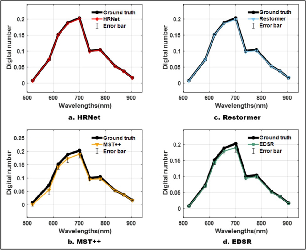
Figure 5. Comparison of average ground truth and reconstructed spectra of egg samples. Spectra extracted from HRNet and Restormer showed the most similarity with their ground truth counterparts, while MST++ struggled to reconstruct most bands.
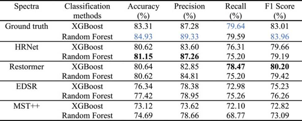
To maintain a common reference basis (i.e., embryo mortality status), only single-day specimens (#102) were used to gauge classification outcomes. Up through day 2, all samples share virtually indistinguishable spectral characteristics, but marked structural and compositional transformations begin emerging from day 3 onward [43]. Accordingly, spectra from day 3 images were used as input for deploying the classification models. It bears emphasizing that merely 9 out of the 102 samples contained non-viable embryos, with fatalities stemming from uncontrolled sources such as genetic anomalies and environmental stressors. To address this severely skewed class distribution within the classification pipeline, the Synthetic Minority Oversampling Technique (SMOTE) was employed to augment the minority class. Using SMOTE, synthetically generated instances representing dead embryos were added to the classification dataset. Stratified 5-fold cross-validation served as the evaluation framework for assessing classifier performance. The aggregated classification outcomes displayed in Table 2 reinforce the spectral reconstruction findings reported earlier.
Table 2. Comparative classification results for predicting live and dead embryos. The blue-colored results indicate the best possible performance obtained from the ground truth, while the bold-colored results show the best possible performance of the reconstruction models.
|
HRNet showed the smallest divergence from the ground truth in spectral recovery. Consistent with this, Table 2 shows that HRNet is the top-performing reconstruction model when paired with Random Forest, achieving accuracies of 81.15%, 87.26%, and 75.20% and an F1 score of 79.19%. Classification metrics derived from Restormer’s reconstructed spectra occupy the second rank among the tested reconstruction models, whereas spectra reconstructed by EDSR and MST++ deliver the weakest classification outcomes.
The reconstruction of hyperspectral imagery holds considerable promise for reshaping agri-vision research by developing affordable, intelligent solutions. This direction opens a pathway to fully harness the power of HSI starting from everyday consumer-grade RGB captures. Recognizing this potential, the present work demonstrates the first hyperspectral image reconstruction for the early prediction of chick embryo mortality during the incubation window. The study deployed HRNET, MST++, Restormer, and EDSR algorithms to recover hyperspectral information from ordinary RGB imagery. With MRAE, RMSE, and PSNR values of 0.0955, 0.0159, and 36.79 dB, respectively, the HRNet model outperformed all other reconstruction techniques evaluated, demonstrating a strong correspondence with both the ground-truth images and their spectra. Building on this, XGBoost and Random Forest classifiers were employed to separate viable from non-viable embryo samples using spectra drawn from the reconstructed images. Consistent with the observed trends in reconstruction quality, the Random Forest model trained on HRNet-reconstructed spectra achieved the strongest classification performance. These promising outcomes encourage further exploration into cost-conscious solutions for hatchery management, propelling the sector toward a more productive, sustainable future aligned with Industry 4.0 principles.
This work was supported by the USDA National Institute of Food and Agriculture, Award # 2023–67015–39154.
None
None
I, Mohammed Kamruzzaman, consciously assure that for the manuscript “Hyperspectral Image Reconstruction for Predicting Chick Embryo Mortality Towards Advancing Egg and Hatchery Industry,” the following is fulfilled:
1) This material is the authors’ own original work, which has not been previously published elsewhere.
2) The paper is not currently being considered for publication elsewhere.
3) The paper reflects the authors’ own research and analysis truthfully and completely.
4) The paper properly credits the meaningful contributions of co-authors and co-researchers.
5) The results are appropriately placed in the context of prior and existing research.
6) All sources used are properly disclosed (correct citation).
7) All authors have been personally and actively involved in substantial work leading to the paper and will take public responsibility for its content.
8) This study followed the animal experiment protocol (protocol #: 22224) approved by the Office of the Vice-Chancellor for Research IACUC, the University of Illinois Urbana-Champaign (UIUC).
Open Access The author(s) retain copyright. This article is licensed under the Creative Commons Attribution-NonCommercial-ShareAlike 4.0 International License. It may be shared and adapted for non-commercial purposes with appropriate attribution, an indication of changes, and distribution of adaptations under the same license. Third-party material may be subject to separate terms identified in its credit line. View the license at https://creativecommons.org/licenses/by-nc-sa/4.0/.
 Agro Intelligence Research Press